See my Science: Laminin:LARGE glycan structure reveals the molecular basis of muscular dystrophy.

August 15, 2016

Laminins are interesting proteins, which is just as well, because they are what I am working on at the moment.

Laminins are large heterotrimeric1 proteins that reside in the extracellular matrix (ECM – the stuff outside cells). If you were to google Laminin, you’d find some stuff about the protein, and a whole mess of stuff about it looking like a crucifix, and how Laminin somehow therefore proves the existence of god and/or intelligent design. It is hopefully clear to you, dear reader, that this is utter nonsense, for 2 reasons:

  1. The diagrams of laminin are exactly that – DIAGRAMS. Cartoons drawn by scientists to make describing these large complex molecules easier. Cartoons of Bugs Bunny are not proof of walking, talking, wise-cracking rabbits;
  2. The pictures (mostly rotary shadowed electron micrographs) of intact laminin are 2 dimensional representations of flexible 3-dimensional molecules, (see fig 1). They don’t look like crucifixes, because they have 3 dimensions – they probably look more like palm trees.
LAMININ, YO.

Fig 1. A Laminin electron micrograph.

This is first protein I’ve worked on that has its own page at Snopes.com, which is quite an achievement.
Right, now I’ve got this curious case of molecular pareidolia off my chest, let’s crack on with why Laminin is pretty damn interesting all by itself.

What Laminins do.

Laminin is a principal component in extracellular structures called ‘basement membranes’ (not the same as lipid/plasma membranes, which you might have heard about in high school biology). Basement membranes are important structures that incorporate Laminins, collagens and other ECM proteins to form protein barriers that separate different tissues layers.

Each Laminin molecule is actually 3 different protein chains attached to each other. Because scientists are exceptionally inventive and imaginative, these three proteins are called Laminin alpha, beta and gamma chains respectively.

They fit together a bit like this (fig 2):

LOOK! IT'S A CRUCIFIX!

Fig 2: Schematic of a Laminin molecule.
Taken from Hohenester & Yurchenco, 2013 http://www.ncbi.nlm.nih.gov/pubmed/23076216

One end of each of the three proteins (the palm tree fronds / the animo-terminus) has an LN-domain (marked LN), and this is involved in forming the basement membrane itself. An LN domain from 3 different Laminin molecules come together to form a sheet like network as shown in fig 3.

Fig 3: How Laminins form a sheet like structure… Taken from Hohenester & Yurchenco, 2013 http://www.ncbi.nlm.nih.gov/pubmed/23076216

This sheet needs to be anchored to the surface of cells. Happily, at the other end of the Laminin, (the base of the palm tree – marked LG1-3/LG4-5 in Fig 2, and marked with a double arrow in Fig 1) there are two functional units that anchor to the surface of cells in two different ways:

    1. By binding to proteins on the surface of the cell called integrins. This happens through the LG1-3 region.
    2. By binding to a specific sugar which is attached to a protein called Dystroglycan. This happens through the LG4-5 region.
      THIS IS WHAT THIS BLOG POST IS ALL ABOUT

Dystroglycan

Dystroglycan is an integral membrane protein – it sits within the plasma membrane (not basement membrane) and has bits that protrude into both the extracellular and intracellular spaces. It links the basement membrane (outside the cell) to the cytoskeleton (a protein framework inside the cell) – see Fig 4.

Dystrophin complex From Baresi & Campbell. http://jcs.biologists.org/content/119/2/199

Fig 4: Dystrophin complex links intracellular (bott0m) and extracellular (top) structures. From Baresi & Campbell. http://jcs.biologists.org/content/119/2/199


The extracellular side of Dystroglycan has a unique sugar polymer covalently attached to it. This sugar (called the ‘LARGE glycan’ or ‘Matriglycan’) has so far only been found attached to Dystroglycan until last week had only been detected on one protein – alpha-dystrogylcan. A recent paper by our collaborators – as demonstrated that this sugar can also be found on other proteins, attached via the same sites known to be occupied by other sugar modifications of proteins (such as the glycosaminoglycans, Chrondroitin Sulphate and Heparan Sulphate). The synthesis of the LARGE glycan is also complex, and the whole linkage region has only recently been mapped. Figure 5 shows a summary of what we know so far about the structure of the LARGE glycan and lists the enzymes that are involved in synthesising it.

dystroglycan-synth

Fig 5: How to make LARGE glycan. By the author, information from various papers.

The business end of the LARGE glycan is the -[Glucuronic acid – β1,3 – Xylose α1,3 ] disaccharide polymer – the repeating orange stars and blue/white diamonds on the right of Fig 5. So what the hell is that then?

It is a long chain of alternating Glucuronic acid (a sugar acid similar to Glucose) and Xylose (a sugar that was first isolated from wood, hence Xylose, derived from the greek for wood) units (see Fig 6). The β1,3 & α1,3 refer to how they are connected.  We don’t know quite how long it is yet, or how many modifications there are on each dystroglycan molecule (either 1 or 2). More research required!

Matriglycan repeating unit.

Fig 6: The LARGE glycan repeating unit. By the author, created using chemdraw 14.0

So why do we care about this?

In a nutshell, muscular dystrophy.

If you cannot make:

  • Laminins
  • Dystroglycan
  • Any of the enzymes involved in LARGE glycan synthesis

you get muscular dystrophy. Loss of the genes that encode LARGE glycan synthesis (i.e. the genes shown in Fig 5) lead to a subset of extremely severe muscular dystrophies called “dystroglycanopathies“. They are congenital (inherited) diseases that prevent correct formation of basement membranes. This means that tissues do not form properly, and gives rise to developmental disorders. If we can understand this system, maybe we can fix it?

Our recent work

So, we know what the LARGE glycan is made up of, and we know that it binds to the Laminin G-type (LG) domain 4 of Laminin alpha 2.

What we want to know is how they interact with each other at an atomic level – happily the best tool for this sort of study is X-ray crystallography, which is what I do for a living!

So I expressed, purified2 and crystallised (Fig 7) a fragment of Laminin alpha 2 which contained the LG4 and LG5 domains. I then soaked a tiny (and I mean tiny) amount of the LARGE glycan that our collaborators had synthesised for us into my crystals. Given that when I did this, we had no idea of tightly Laminin alpha 2 bound to the LARGE glycan, this was a bit of a long shot. I setup around 20 different experiments, with different soaking times and concentrations to try and get some ligand into the protein.

Crystals!

Fig 7: Crystals!


I cryocooled the crystals in liquid nitrogen and sent them off to Diamond Light source, where we collect our diffraction data. My remote access data collection shift started at 4am. Oh joy. An early start, plenty of coffee and a taxi to work and I was good to go. However – a drawback of the ligand soaking approach I had to take was that I would have no idea whether or not my crystals had the ligand in until I had processed all the data. I didn’t have the time to process the data as I went along, so the data collection was done using ‘the American method’ – Shoot first, ask questions later.

After my 5-hour shift was up, I grabbed breakfast and more coffee and started to process all my data.

About 5 datasets into my haul of data, I hit the jackpot.

Fig 7

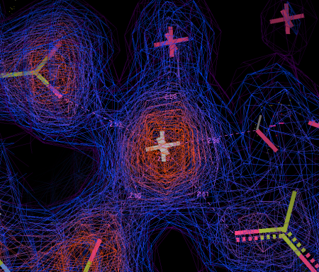
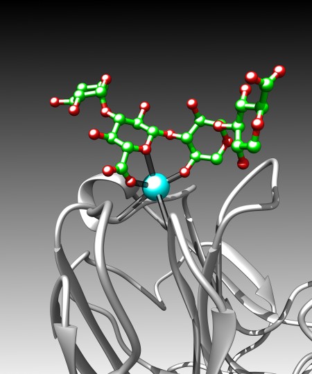
Fig 8a : What the Calcium binding site in LG4 looks like.


Fig 7: Difference density map of SOMETHING.

Fig 8b: Difference density map of SOMETHING.

Above the calcium binding site in LG4, right where it was predicted to be, was a big green blob in my electron density difference map. If you look at Fig 8a, you can see what the empty binding site looks like – this is a very much simplified representation as there is an awful lot going on. The cyan sphere is a calcium ion. The red/green/blue sticks around the calcium show the orientation of the amino acids in the protein that bind to the calcium. The loops and whirls represent other bits of the protein.

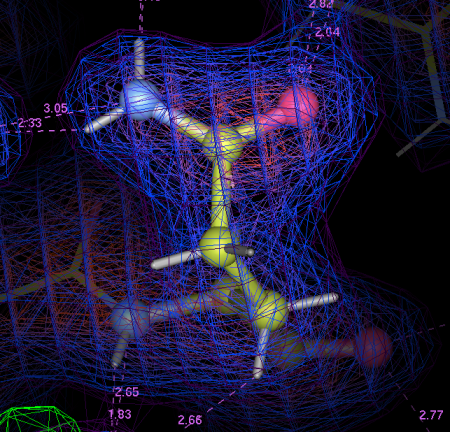
I generated an electron density map that highlights the differences between an empty crystal and a soaked crystals (fig 8b). To generate this map, I simply subtract data from an empty crystal (apo form) from data of a soaked crystals (bound form) – the difference between the two datasets should equal the ligand. Happily, it does! After a bit of building and refining, we end up with the final refined structure of the Laminin:LARGE glycan complex.

Fig 9

Fig 9: THAR SHE BLOWS!

Figure 9 shows this final structure of the ligand with the same density shown in Fig 8, just made transparent.


Fig 10

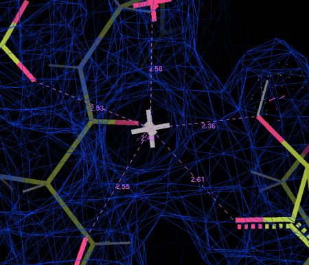
Fig 10: A schematic of important interactions in-between Laminin (red) and the LARGE glycan (black).

When we dive into the binding site and take a look around (Fig 10), we see that all the interactions are between a single glucuronic acid – xylose repeat, even though there are 3 repeats in the sample of LARGE glycan we used. The link between the two sugar rings straddles the calcium ion, forming a sort of chelating or clathrate-type interaction. Oxygen atoms (carrying a net negative charge) interact strongly with the positively charged calcium ion . The carboxylic acid group in the glucuronic acid ring (The O=C=O bit) also pokes into a little positively charged pocket formed by two backbone amino groups (NH). All these interactions combine to form quite a strong interaction for (~0.2µM KD, if anyone is interested) for an interaction with a relatively small interaction surface.

So what have we learned?

We now know exactly how Laminins bind to the LARGE glycan. Let’s be clear – this is not a drug target – we definitely do not want to inhibit this interaction! But this shows us a crucial link in the chain between the cytoskeleton (the internal skeleton of a cell) and the outside world.

We have also seen a really interesting and novel mode of protein-carbohydrate interaction. The Laminin does not recognise individual sugars – it recognises the unique linkage found in the LARGE glycan. Given that there are over 100 proteins in the human genome that contain LG domains, and some of those are also known to bind to the LARGE glycan, our structure provides a paradigm for LG domain – LARGE glycan interactions.

We know that a single disaccharide is sufficient for Laminin to engage (although longer stretches of sugar bind more tightly) and we show that the sugar forms a pseudo-clathrate cage over the calcium in the protein. This is probably why the Laminin-LARGE interaction is ~10x tighter than interactions between proteins and other similar sugars.

Anyway – the paper should be out NOW in Nature Chemical Biology.

A PDF is available here.

The pdb files are here

binding_cartoon_gradient

IMAG0194 copy

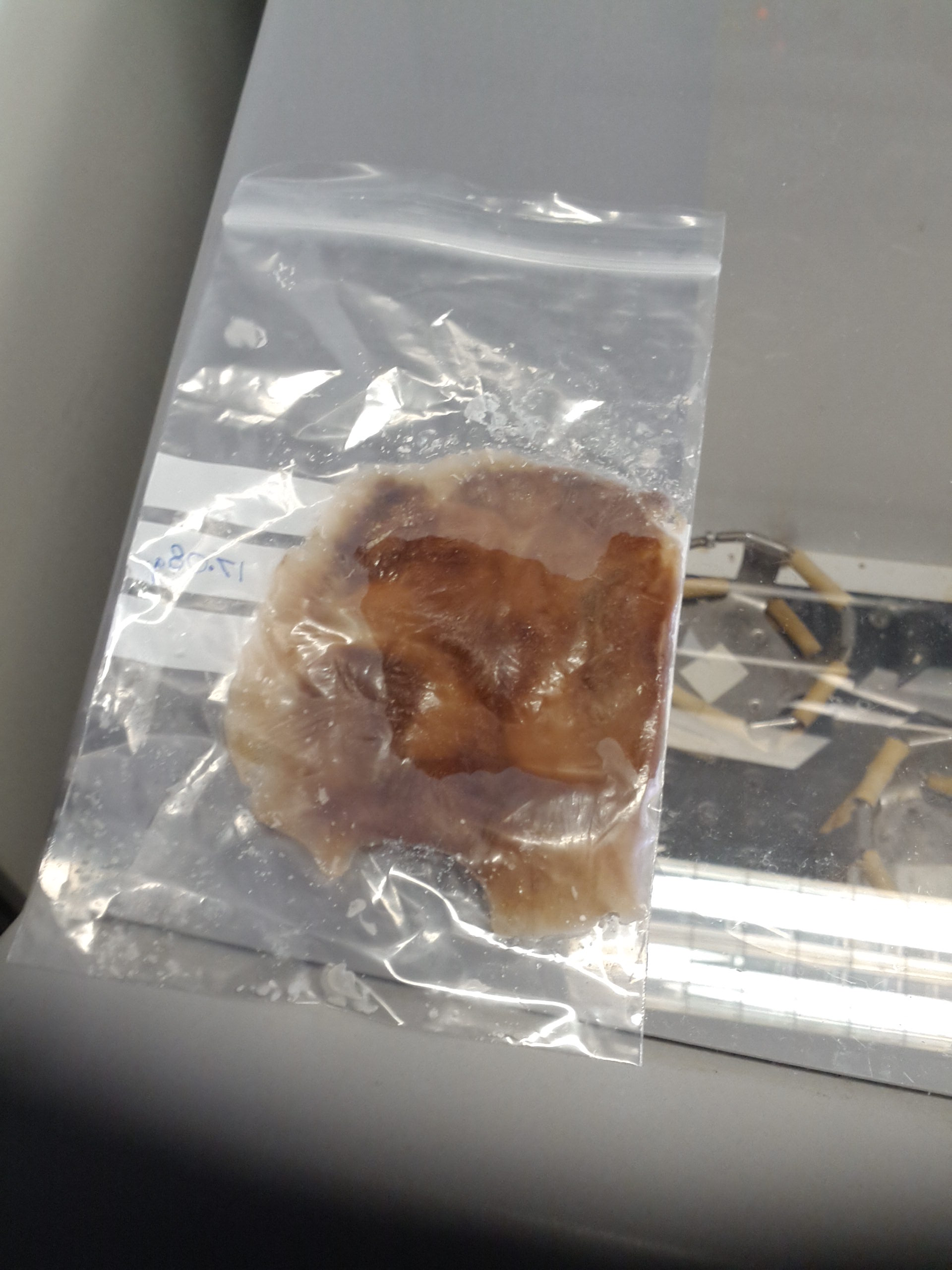
The author’s right forearm.


  1. Hetero – different, trimer – three. Made up of 3 different proteins.
  2. Made in mammalian cells – we give mammalian cells DNA that encodes our protein.

Multi-contoured electron Density maps

December 4, 2015

When wandering around the department, I am struck by how few crystallographers use multiple contoured electron density maps* whilst building. I really don’t understand this: YOU’RE THROWING AWAY INFORMATION PEOPLE!

Even at moderate resolution, the information gained can be invaluable:

  1. Precisely locating a heavy atom in a big blob of electron density. The Atom will sit at the highest point of the map. IF you are using multiple contours, this will be obvious! (figure 1)
  2. Resolving His/Asn/Gln sidechain flips. A bit more prone to noise here – but in terms of electrons, O > N > C. You can easily decide which way around Asn and Gln side chains should point, and often get some help with His side chains as well. (figure 2)
Screenshot 2015-12-04 13.05.44

Figure 1a: THAR SHE BLOWS!

This calcium ion sits RIGHT on the peak in the electron density map. No ambiguity where it lies. A single map contoured at 1 sigma is not helpful.

Screenshot 2015-12-04 13.07.38

Figure 1b: Nope. Not helpful.


Screenshot 2015-12-04 12.54.32

Figure 2: Gln sidechain flips, made easy…

The increased electron density  on the right hand side here indicate that this Gln residue is the correct way around. Again, a single map contoured at 1 sigma is no use here.

I realise that there are other ways to achieve what I have described, but when you are building your models, saving time and making things easier is hugely helpful. I find using multi-contouring incredibly helpful.

* multi-contours shown here are made using the “Multi-chicken” command in COOT (extensions>maps>multi-chicken). Multi-chicken creates 10 maps contoured at 1,1.5,2, 2.5, etc sigma. I find the default setting is a tad dark so I use “brighten maps” (extensions>maps>brighten maps) a couple of times to sort that out. I then contour the original map at 0.7sigma (depending upon noise) and make it really deep purple.

All screenshots made with COOT.


Outright deception.

June 6, 2014

Sorry – Homeopathy again.

EDIT – There is an update that follows the original post

Those who have ventured “below the line” on articles regarding homeopathy will perhaps have come across “Dr” Nancy Malik, a prolific pro-homeopathy zealot who can be typified by her chronic inability to read and assess the articles she is touting as showing that her beloved modality is anything other than water.

In the not too distance past, Ms Malik had a “knol” – a Google hosted blog where she collected all the scientific papers that she thought showed homeopathy worked. I assessed that here.

(Spoilers: there’s nothing in it.)

Google pulled the plug on Knol, and Ms Malik migrated her site to WordPress.
A few months ago, she got the site HONcode certified. HONcode is an independent organisation that promotes and certifies websites that they deem as giving reliable healthcare information. Obviously, I found it odd that HONcode (who seek to abide by the tenets evidence based medicine) would certify a homeopathy site. I raised this with them, and encouraged others to do so. After a brief e-mail conversation, HONcode wisely chose to suspend Ms Malik’s certification, pending a review. The HONcode logo on her site and the verification link (supplied by HONcode) changed to reflect this – now showing a ‘men at work’ sign with a red “ReExam” warning.

Both myself and Alan Henness asked Ms Malik when she might be altering her website to take this into account. This morning, after several weeks gentle prompting, we got a reply…

image

Her website HAD been changed!

image

Or had it? The HONcode logo still has the red “ReExam” logo, but when one clicked on the verify link… Something magical happened!

Rather than the usual link to the HONcode site with further information about the certification…

The real HONcode site

The real HONcode site

… one is directed to an image of the original certificate posted on Ms Malik’s Google+ site in October 2013.

image

The G+ image

Here is a freezepage link to Ms Malik’s WordPress site as of this morning, complete with link to her G+ site.

This is a clear and unambiguous attempt to dupe the unwary into thinking that she retains HONcode certification. Happily it was so laughably crude that even my pre-coffee eyes at 6:50 spotted it.

The genuine HONcode ceritificate for Ms Malik’s site is here.

EDIT: I’ve added some links & an image of the genuine HONcode certificate.


UPDATE: Alan Henness was the first of us to illicit a response from HONcode, who appear to confirm that they have rescinded their certification for Ms Malik’s site. Perhaps we might speculate that her sudden action on this issue (after 6 weeks of nought but silence on the matter)  might have been precipitated by HONcode informing her of their decision?

UPDATE 2: HONcode have now removed Maliks certificate – the link to here genuine certificate now looks like this:

Screenshot 2014-07-10 at 22.54.30


BUG SPLATS!

February 28, 2014

A quick technical note/tip for folks doing recombinant protein expression in E.coli


When doing large-ish scale expression of proteins in E.coli, it is common to freeze the cell pellet post-harvesting and prior to cell lysis and protein purification. Typically this is achieved by re-suspending the cell pellet in a small about of Luria Broth or PBS and then re-pelleting the cells in a 50ml falcon tube, and freezing them at -80ºC.

Whilst this is convenient, the down side of this is that it introduces another centrifugation step into your harvesting protocol, and a solid lump of cell paste can take a while to thaw out fully, and can be difficult to get completely homogenous and lump-free prior to whatever lysis technique you choose to use.

A colleague of mine who recently joined the lab from AstraZeneca (after they closed much of the Alderley Park research facility) has brought with him an ingenious way of speeding both harvesting and thawing out considerably: BUG SPLATS.

Poo in a bag?

Revolutionary bacteria pellet freezing protocol!

Rather than re-suspend your pellet in LB/PBS and re-pellet it, just scoop it out with a spatula (or similar) and place the bug pellet into a small (~10x20cm) press-lock bag.

Collect all the pellet into the bottom of the bag and then smooth it out so that your bug splat is nice and thin (<5mm is ideal), and stick it in the freezer.

The thin bug splat will freeze faster than a pellet in the bottom of a falcon tube.

It will also thaw out much faster. For thawing – snap the brittle, frozen bug splat into small chunks, and empty them into a suitable container (50ml falcon or similar), and add the required amount of lysis/sonication buffer. It should thaw out rapidly, and it should be easier to obtain a smooth, homogenous solution before lysis than a large ~25ml lump of frozen paste in the bottom of a falcon tube.

As the pellet is brittle, you can also snap smaller piece off if you don’t want to thaw the whole thing out.

So there you go – a ludicrously simple way of saving time in the lab, and making the freeze/thaw process faster, which is (anecdotally) better for your protein.


The week in Structures – 8th May 2013 #PDB

May 8, 2013

Week #2: Trying to jumpstart our immune system to fight off HIV. 


Fighting the good fight against HIV.

In this weeks PDB releases, we see a couple of HIV-antibody structures, continuing the work of the Kwong and Mascola labs in the NIH. In 2011, these labs released a paper (published in Science) studying various antibodies that recognises and neutralise HIV-1. If you can recall your high school biology classes, antibody recognition of an invading pathogen is the first step in activating the immune system that hopefully results in successfully fighting off the infection. This work has been further developed with papers this year in Cell and Nature – Prolific and important work!

The structure shown below contains the “FAb fragment” (gold & bronze cartoon) of the antibody bound to gp120 (the big blue blob).

Antibody recognition of HIV gp120 - a surface marker of the HIV virus. After PDB # 4JB9

Antibody recognition of HIV gp120 – a surface marker of the HIV virus. After PDB # 4JB9

Glycoprotein 120 (gp120) is a component of the HIV surface spikes – which recognise a range of different molecules on the surface of our cells and initiate infection. It is because of the this that gp120 is an attractive vaccine candidate – however, recombinant gp120 has failed deliver results in clinical trials, and it is hoped that by studying antibody-gp120 interactions and perhaps by designing a novel gp120-dervied antigen, an effective HIV vaccine can be developed.

Presumably the authors discuss the relevance of their findings in the paper (not released at the time of writing here) in the context of HIV treatment and rational vaccine design. (PDB codes 4JB9 and 4J6R). (EDIT: Other work in the same edition of Science discuss similar topics, here and an article “Rational HIV Immunogen design to targets specific germline B-cell receptors.”)

Other structures of note this week at the excellently named “star domain of quaking protein in complex with RNA” and some work looking at the mechanism by which angiopoietin 1 and 2 interact with the Tie2 receptor tyrosine kinase ([paper] | [PDB] ), which might lead to therapeutics that prevent tumours growing their own blood supplies (angiogenesis blockers).


On branded drugs.

May 5, 2013

I went shopping yesterday…


Anti-allergy meds

On the left, a box of 14 generic allergy relief tablets, each containing 10mg Cetirizine HCl and Lactose – costing £1.

On the right, a box of 14 branded allergy relief tablets, each containing 10mg Cetirizine HCl and Lactose – costing £5.67. In a sale. Down from £7.57.

The active ingredients are  identical. The evidence for the efficacy of the active ingredients is identical. So how can companies justify charging 7.5 times more? I understand and acknowledge that effective marketing and other psychological factors might lead to a more effective placebo component of any clinical effect [1][2] – but a 7.5 fold increase in effectiveness?

The chemical structure of cetirizine.

The chemical structure of cetirizine. (Photo credit: Wikipedia)

If we assume that the sellers make some sort of profit on the generic, then someone must be making a huge profit on the branded anti-allergy meds. Which seems a little immoral. If you can offer people relief for 7.1p per day, why charge as much as 54p per day?

Whether or not this pricing is down to the pharmaceutical industry I cannot be sure – but big pharma don’t have the greatest public image at the moment – and examples like this sat on the shelf of your local supermarket perhaps serve as another example of why.


The week in Structures – 1st May 2013 #PDB

May 1, 2013

The first in a (hopefully) weekly series of reviews of interesting structures in latest PDB release**.


High-resolution* Cryo-EM models of Human and Drosophilia Ribosomes!

Proteins are the major molecular players in life as we know it – they are the chemists, the engineers, the messengers and the defenders of life. They make new chemical compounds that we need, they break old compounds down so that we might re-use them – they transmit messages between different cells and tissues and help identify invading pathogens. In short – proteins do pretty much everything, and as such are the subject of intense study and scrutiny.

What better subject for the first of my PDB release highlight posts, than the Ribosome – a vast (at least at a molecular level) and ancient molecular machines that are responsible for the synthesis of proteins in our cells. The (perhaps outdated) “central dogma” of biology is that DNA is transcribed to mRNA, which is translated into protein, and the proteins then go and do everything. Anger et al have released 5Å resolution Cryo-EM-derived models of human and drosophila (Fruit Fly) ribosomes – they are so vast that they have to be split across several PDB files each…

Human (3J3A, 3J3D, 3J3B, 3J3F)

Drosophila (3J38, 3J3C, 3J39, 3J3E)

We're gonna need a bigger boat.

… and so complex as to defy creation of a clear and crisp picture.

The Paper concerning these is due to be published in Nature, but is not available at the time of writing (EDIT – Now it is – here (£)). Presumably, along with the methods  employed in what must be a massive modelling task, there will be some discussion of the differences between these two eukaryotic ribosomes, and the prokaryotic ribosome structures that were the subject of the 2009 Nobel Prize for Chemistry.

Other highlights in this weeks crop of 287 PDB releases include yet more BACE-inhibitor complexes (potential lead compounds for Altzheimer‘s treatments) and CDK2 and CDK8 -inhibitor complexes, which may be lead to novel anti-cancer therapies.


*ok – 5Å might not be great for crystallography, but for our Cryo-EM-based friends, this is pushing the limits and is clearly the result of a great deal of hard work and a massive amount of particle picking.

** The PDB is the “Protein Data Bank” – all structures of proteins/DNA and related molecules are deposited into the PDB and then made available to everyone (for free) prior to or at the time of publication of the paper that describes the work pertaining to the structure. The PDB issues new releases every Wednesday morning (UK time).


Like science?

April 30, 2013

Like science and the progress and prosperity it brings the UK? Then sign the pants off this bad boy. http://ow.ly/kyyQ7 #sivpoint8


POP QUIZ – What’s this?

April 16, 2013
Erm...

What the hell is this thing?

A little game – What the hell is this thing? Take a guess in the comments below – the red blobs are me deleting labels that might have given it away.

The prize is kudos, the admiration of your peers, and a life time subscription to this blog. 😉


#QEDcon 2013 ISS passes

April 9, 2013

Information shamelessly lifted from meteorwatch.org and metcheck.com.

Friday Night (12th)

  • 21:43-21:48 – “Incredibly Bright” – cloud cover 23%

Saturday Night (13th)

  • 20:52-20:59 – “Incredibly Bright” – cloud cover 99% 😦
  • 22:29-22:32 – “Incredibly Bright / Short pass” – cloud cover 100% 😦

Sunday Night (14th) (for the stragglers)

  • 21:39-21:44 – “Incredibly Bright” – cloud cover 94% 😦

So… Friday night it is then, caveats about weather forecasting notwithstanding. If we can drag ourselves out of the bar for 5 minutes.